
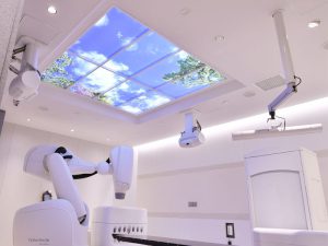
 Cyber Knife M6
The Cyber Knife Robotic Radiosurgery System is a noninvasive treatment which delivers beams of high dose radiation to tumors in various parts of the body. The Cyber Knife System can capture the exact location of the tumor using x-ray images, and determine the size and the shape of the tumor with a CT scan. It delivers highly focused beams of radiation with a flexible robotic arm, which allows physicians to treat areas of the body that cannot be treated with other radiology techniques.
 TomoTherapy
TomoTherapy utilizes 3D image to capture the position and the size of the tumor, which enables the delivery of precise radiation therapy. The TomoTherapy treatment system can deliver the radiation therapy continuously, and adjust the intensity of the radiation beams throughout the session to minimize the radiation which reach healthy surrounding tissues and organs. An advanced technology of TomoTherapy treatment system can deliver radiation therapy continuously to multiple cancers at once, and even to lesions located in unreachable places by other systems. The procedure lasts about 10 to 20 minutes.
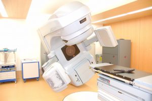 Linac
Linac
Linear Accelerator (LINAC) therapy is the most advanced radiation treatment for all cancers. The LINAC technology provides high energy x-ray with high accuracy to targeted tumor, sparing healthy surrounding tissues and organs.
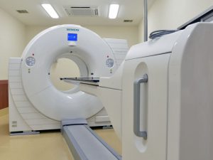
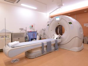 Computed tomography (CT)
A computed tomography (CT) scan combines a series of X-ray images taken from various angles to examine inside the body from multiple angles to detect conditions in organs and body parts such as brain, chest, abdomen, joints, and pelvis.
CT can produce higher quality images than a X-ray within a short amount of time. Most examinations will take just a few minutes to complete.
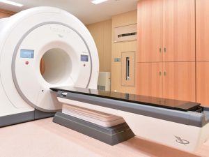
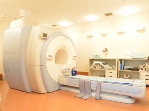 Magnetic Resonance Imaging (MRI)
A magnetic resonance imaging (MRI) is similar to a CT scan; however it does not use X-rays like a CT scan. Instead, it uses a strong magnetic field and radio waves to produce very detailed images of internal body structure, such as organs and bone. The blood vessels can also be examined with a special kind of the method, called magnetic resonance angiography (MRA). A MRI is superior in detecting tumor and lesions in soft tissues over a CT scan, however, most MRI examinations will take longer than fifteen minutes.
 Nuclear Medicine (NM)
Nuclear medicine uses very small amounts of nuclear medicine (radioactive materials), or radiopharmaceuticals, to examine organs and tissue structure as well as function. Nuclear medicine is noninvasive except radioactive material is injected through an intravenous line. Nuclear Medicine allows healthcare professionals to observe the progress and early stages of diseases and how the disease is affecting the function of organs.
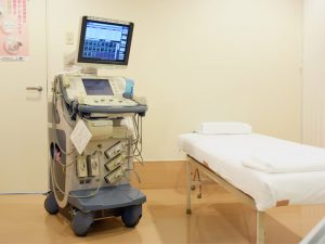 Ultrasound diagnostic device(US)
Ultrasound (US) diagnostic device uses special sound waves to produce pictures of body. Because Ultrasound captures real-time images of internal organs, heart, tendon, and fetus, doctors and other healthcare professionals can view movements of internal organs.
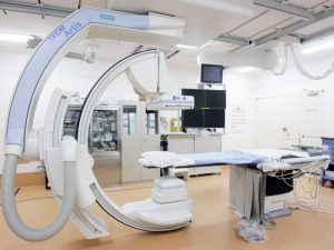 Interventional Radiology (IVR)
Interventional Radiology (IVR) provides minimally invasive treatment and diagnosis using x-ray image guidance.
A catheter is inserted through a small incision to deliver contrast substance and treatment agents for various conditions such as aneurysms and atherosclerosis.
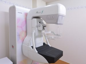 Digital Mammography
Mammography is the best screening x-ray technology for detecting early stage breast cancer and diagnosing breast disease in women. Digital mammography is more effective at differentiating tumor from dense fibrocystic breast tissue by allowing physicians to manipulate the digital image.
 Gastrointestinal Endoscope
An endoscopy is a medical procedure to examine digestive tract like esophagus, stomach and intestine with an instrument called an endoscope. Endoscopes are also used for surgeries to remove both cancerous and noncancerous tumors from digestive tract.